
Muscle development may be guided by more than genes and chemical signals. A new study from the Max-Planck-Zentrum für Physik und Medizin finds that physical forces inside developing tissue help determine where muscle cells join together—a discovery that could reshape how researchers think about growth, regeneration, and repair.
As first reported by Phys.org, the research examines myoblasts, precursor cells that merge into elongated structures called myotubes before developing into mature skeletal muscle fibers. Rather than fusing evenly across a sheet of cells, the myoblasts preferentially came together in localized regions where mechanical stress was especially high.
The team, led by researchers including MPZPM postdoctoral researcher Dr. Sushil Dubey and Prof. Benoît Ladoux, combined an in-vitro chicken myoblast model with live-cell imaging and force-mapping techniques. Their findings, published in Nature Communications as “Extracellular-matrix mediated stresses spatially bias myoblast fusion and myotube growth,” identify the extracellular matrix—the material surrounding cells—as an active participant in the process.
The researchers describe these high-stress areas as mechanical hotspots. In the developing cell layer, organizational irregularities known as topological defects create regions of elevated compressive force. Those sites become more likely locations for fusion, suggesting that the physical layout of a tissue can influence when and where the molecular machinery of muscle formation is used.
The process also appears to reinforce itself. Once myoblasts fuse, the resulting myotubes become more aligned, changing the tissue’s overall organization and increasing local stresses. Cells simultaneously remodel the extracellular matrix, helping preserve the force patterns that guide later fusion events. In effect, developing muscle behaves less like a collection of isolated cells and more like an active material whose structure and forces evolve together.
That distinction matters because understanding muscle formation requires more than identifying the molecular switches that permit cell fusion. The MPZPM findings suggest that the surrounding mechanical environment helps decide where fusion is most likely to happen. The work could eventually provide useful context for studying impaired muscle development, tissue regeneration, and disorders in which repair fails, although the experiments remain a laboratory cell-model study rather than a clinical treatment.
The paper gives researchers a framework for investigating how physical forces interact with biochemical signals during tissue formation. The next challenge will be determining whether the same stress-guided behavior operates in more complex animal models and injured human muscle. For now, the study adds a literal dimension to the biology of strength: muscle architecture is partly built by the forces generated as cells organize themselves.
Image Credits
In-Article Image Credits
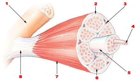
Skeletal muscle tissue via Wikimedia Commons by National Institutes of Health with usage type - Public DomainFeatured Image Credit
Skeletal muscle tissue via Wikimedia Commons by National Institutes of Health with usage type - Public Domain


